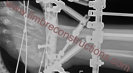
Further alignment improvements observed on X-ray and clinically. New prescription issued to fine-tune valgus, equinus, and medial translation.
Part 6 (23 November 2018) – Aiming in Tibio-Calcaneal Fusion Using Fine Wire Frame
Further alignment improvements observed on X-ray and clinically. New prescription issued to fine-tune valgus, equinus, and medial translation.
First correction completed with improved alignment visible on X-rays. Progressing as planned.
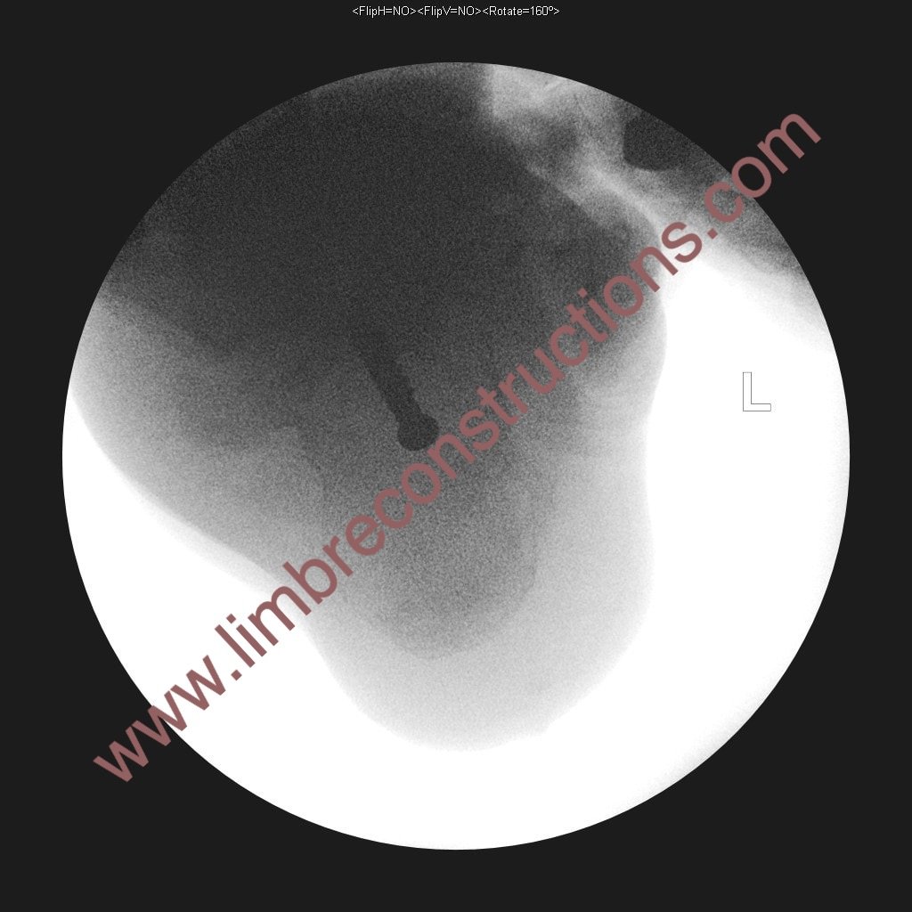
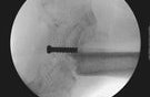
X-rays show the screw is clear in the AP view but less visible in the lateral view, which was never the main issue.
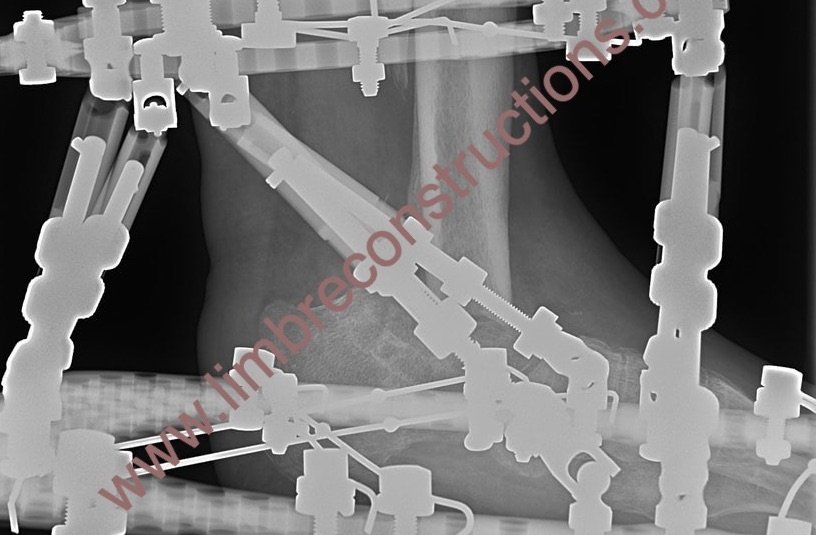
During revision surgery, a 6.5mm cancellous cannulated screw was carefully positioned to guide docking, though the optimal tibial docking site remains uncertain—whether on the calcaneum alone or both the calcaneum and navicular. A new foot plate was attached, but the docking site was not modified per standard procedure.
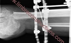
Following resection of the distal tibia and talus due to chronic osteomyelitis, bone transport using a fine wire frame was performed. However, misalignment at the docking site became evident only after frame removal. A review of similar cases suggests that the commonly used “dynamisation” period may not be a reliable test for confirming bony healing.
Achieving accurate alignment in tibio-calcaneal fusion using a fine wire frame is a significant challenge. While past successes have relied on luck, recent failures prompted a reassessment of targeting methods. With limited imaging options, a 6.5mm cannulated cancellous screw is now being explored as a surgical target marker to improve precision.