Part 3 – 4 August 2015
One week after we started with the compression of the fracture site X -ray suggests that fragments are traveling in the planned direction. Speed of compression is 2mm per day, when distraction of distal corticotomy is 1 mm per day. Leg is still shorter for around 1 inch but will get shorter in next few weeks for more than 2 inches altogether. This is my current estimate, but I don’t know how far into the mid fragment we will be able to get the proximal fragment. More compression we can get, the better.
Corticotomy side is distracted for another 7mm (14mm altogether) and good bone regenerate is visible. All pin sites are dry including proximal half pin which is HA coated. Today we changed one strut, another one is due in next few days (as per TSF prescription).
Ap and lateral view X-rays are suggesting good apposition of the proximal fragments and good callus formation in the level of corticotomy.

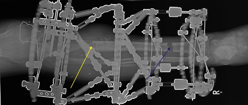
AP view after compression of the proximal fracture started (Yellow arrow) and distraction of the distal rortirotomy progressed to around 15mm (Blue arrow).

Lateral view after compression of the proximal fracture started (Yellow arrow) and distraction of the distal corticotomy progressed to around 14mm (Blue arrow).
Plan:
- Continue with compression of the fracture site until struts get realy tight (we never use spanner to turn struts).
- Continue with distraction of the corticotomy site with the speed of 1mm/day.
- Weight bearing as tolerated, physiotherapy for lower leg; knee and ankle joint.
- Follow up in one week with another X-ray on arrival to check proximal docking.