Part 2 (23 October 2018) – Aiming in Tibio-Calcaneal Fusion Using Fine Wire Frame

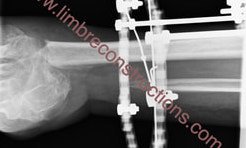
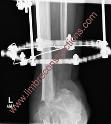
Because of the chronic osteomyelitis of the distal tibia and talus, distal tibia and talus were resected. Proximal tibia corticotomy and bone transport using fine wire frame filled the bone defect, but I missed the docking site with the frame. Not good.
In my defence, “dynamisation” period was uneventful and the failure only became obvious after the foot plate was removed.
At this point I looked at other patients where docking site fusion failed after removal of the frame. Surprisingly, they all completed 4 weeks of dynamisation without any problem. My conclusion so far is that whilst dynamisation sounds like a very sensible option, it is very likely not helpful in assessing bony healing. Until we get further evidence we will not use dynamisation as a docking site union confirmation test.


Author: Matija Krkovic
Website: https://www.limbreconstructions.com/