Limb Reconstructions > Blog > Clinical Blog > Complex Lower Leg Injury > Part 15 (5 May 2016) – Complex Lower Leg Injury with Significant Injury to the Knee Joint and Extensor Mechanism
Part 15 (5 May 2016) – Complex Lower Leg Injury with Significant Injury to the Knee Joint and Extensor Mechanism
Part 15 – 5 June 2016
One of the proximal wires broke and was removed today. The rest of the wires still solid and pin sites clean. Knee started to grind and made noises. Unfortunately something we expected.
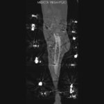
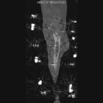
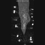
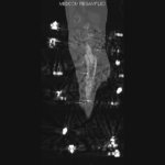
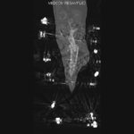
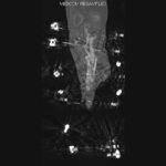
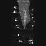
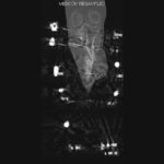
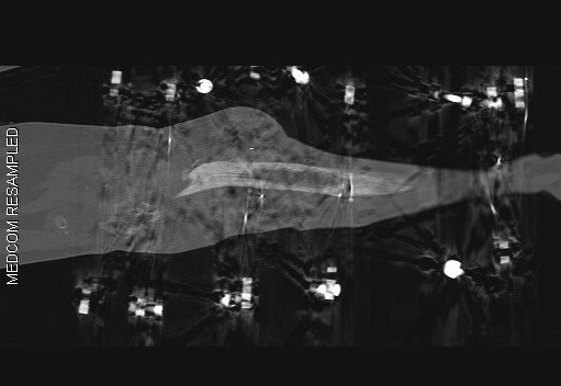
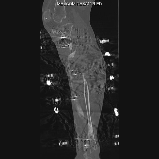
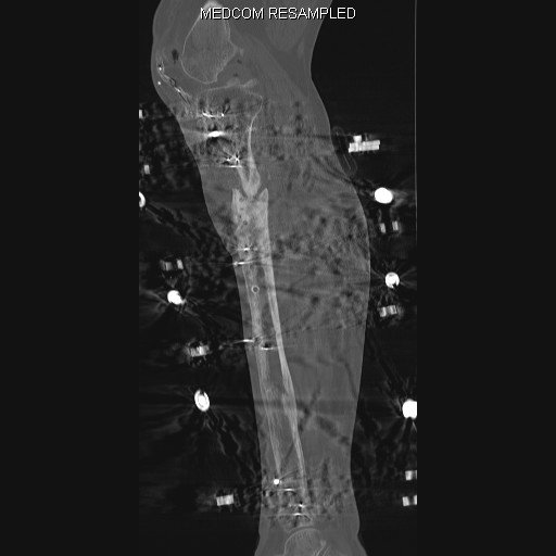
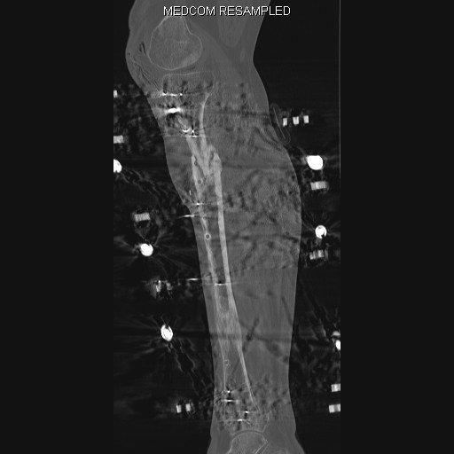
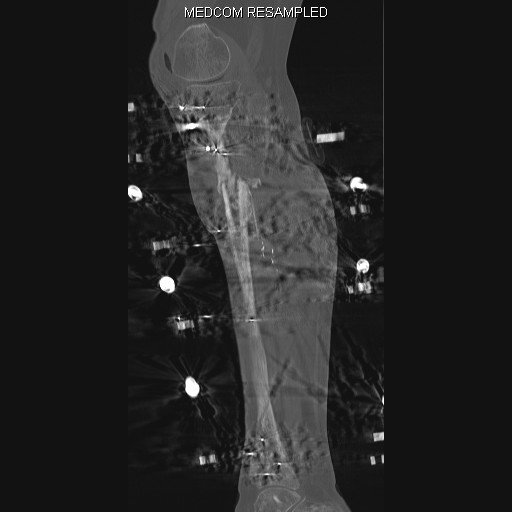
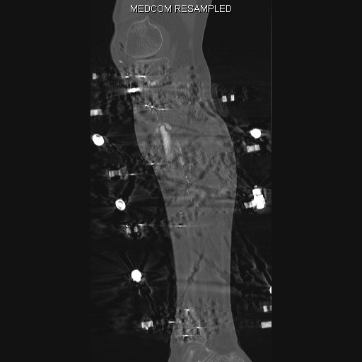
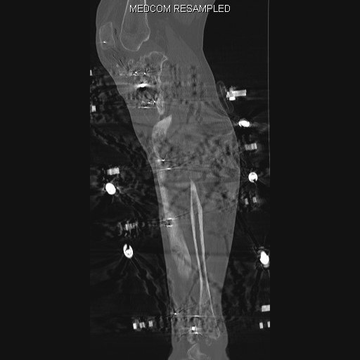
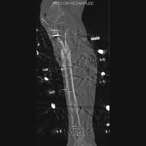
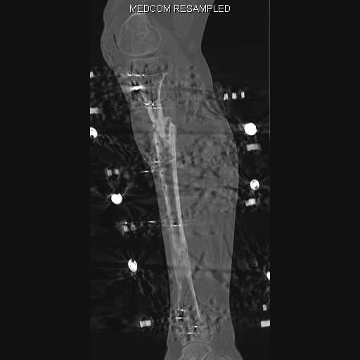
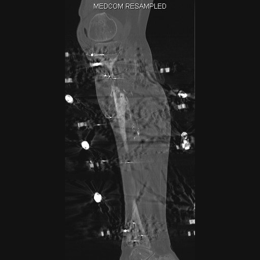
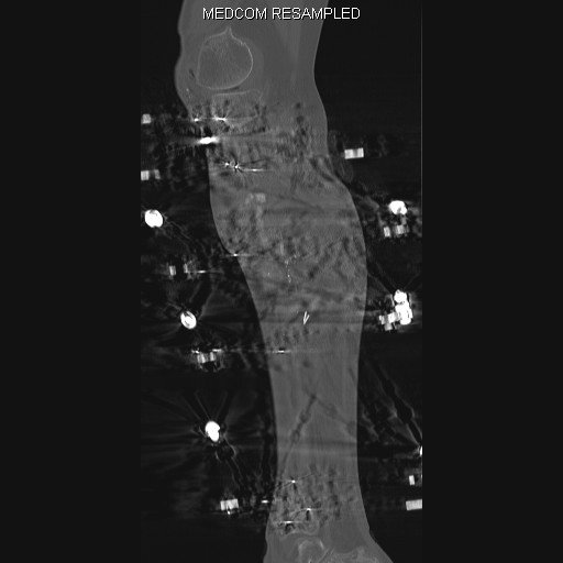
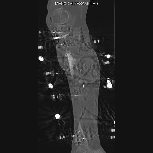
CT scan confirmed good biological response on the fracture side but no clear union as yet. Regenerate is almost completely ossified. No other untoward signs.
CT scan – Coronal reconstruction






Plan:
- Continue with the treatment
- Frame will have to remain on the leg for another 6 months
- Will not remove part of the frame (might compromise stability)
- Further follow up 6-8 weeks
bone healing CT scan external fixation fracture healing frame removal gait improvement limb reconstruction orthopedic surgery physiotherapy post-surgical recovery Taylor Spatial Frame tibial alignment weight-bearing

Author: Matija Krkovic
Website: https://www.limbreconstructions.com/
I am a consultant orthopaedic trauma surgeon working at Addenbrooke's hospital, Cambridge University Hospitals NHS Foundation Trust.
Search
Categories
Recent Comments
No comments to show.
