Limb Reconstructions > Blog > Clinical Blog > Complex Lower Leg Injury > Part 12 (1 March 2016) – Complex Lower Leg Injury with Significant Injury to the Knee Joint and Extensor Mechanism
Part 12 (1 March 2016) – Complex Lower Leg Injury with Significant Injury to the Knee Joint and Extensor Mechanism

Part 12 – 1 March 2016
Walking with one stick. Knee feels more stable and reliable. Pin sites are still OK.
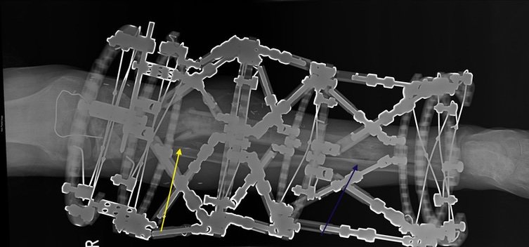
X-ray shows excellent consolidation of the regenerate (blue arrow) and further healing of the fracture (yellow arrow). Possible sinus mentioned previously has not reoccured.


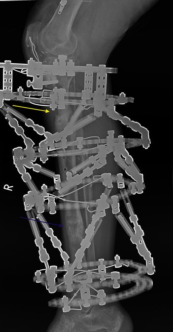
Ap and lat radiographs of the tibia. Good alignment and further progress in the fracture union (yellow arrow) and further maturation of the regenerate (blue arrow).
Plan:
- Continue with physio and exercises
- Weight bearing as tolerated
- Follow up in 6 weeks
bone healing external fixation fracture union gait improvement limb reconstruction orthopedic surgery physiotherapy post-surgical recovery Taylor Spatial Frame tibial alignment weight-bearing

Author: Matija Krkovic
Website: https://www.limbreconstructions.com/
I am a consultant orthopaedic trauma surgeon working at Addenbrooke's hospital, Cambridge University Hospitals NHS Foundation Trust.
Search
Categories
Recent Comments
No comments to show.