Part 6 – 11 Spetember 2015
I was on a holiday, therefore a bigger gap. But everything was under control.
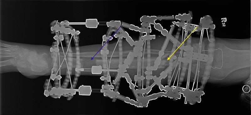
Proximal fracture (yellow arrow) was translated and compressed further with some visual effect on the latest X-rays and corticotomy gained further length (blue arrow). Almost reached the length of the leg as per original plan. Another inch or so to go. Had to change clickers for longer ones (will be visible on the next X-ray next time) and longer threaded bars to allow further lengthening. No obvious infection around pin sites.

AP radiograph of the right lower leg post compression of the proximal fracture (yellow arrow) and distraction osteogenesis distal tibia (blue arrow).

Lateral radiograph of the right lower leg post compression of the proximal fracture (yellow arrow) and distraction osteogenesis distal tibia (blue arrow).
Plan:
- Continue with distraction until the same length is achieved. We might over lengthen it a bit and than compress to add to consolidation time.
- Continue on exercising the ankle to improve range of movement lost due to the lengthening
- Proximal fracture is compressed and will leave it for now as it is (struts are tight and cannot be moved without using a tool)
- Careful monitoring regarding possible osteomyelitis