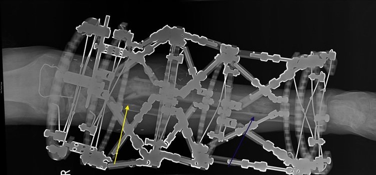
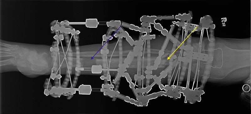
The patient is now walking with one stick, with improved knee stability and no pin site issues. X-rays show excellent bone regeneration and further fracture healing. Continued physiotherapy and weight-bearing as tolerated are planned.
The final TSF prescription has been completed successfully, with improved tibial alignment. The patient is walking unaided for short distances, though ankle dorsiflexion remains a challenge. Continued physiotherapy and monitoring of a suspected low-grade infection are planned.
A recent CT scan shows promising signs of healing, with good callus formation and successful alignment of the proximal fragment. Adjustments to the TSF prescription will further refine leg positioning.
Leg alignment has been successfully corrected, improving gait stability. Early signs of callus formation at the proximal fracture site are promising, with a CT scan scheduled to confirm progress.
The healing process continues as expected, with significant improvement in swelling and early callus formation. However, mild valgus alignment has been noted, and potential corrective action is being considered to optimise long-term function.
Distraction osteogenesis is complete, with both legs now equal in length. Encouraging signs of healing are visible on X-rays, including unexpected early callus formation at the fracture site. The focus now shifts to rehabilitation and ongoing monitoring.
Steady progress continues with further lengthening nearing the planned goal. Compression at the proximal fracture site is holding, with no signs of infection. Adjustments made to allow additional distraction.
Significant progress as the proximal fragment reaches the docking point. A minor deformity correction is planned, with continued distraction at a reduced rate to minimise stress on the ankle.
Progress continues in this complex limb reconstruction case using the Ilizarov apparatus. The tibial corticotomy has been completed, and bone lengthening is underway with visible regenerate formation. The proximal fracture has been disimpacted, aligned, and prepared for docking into the medullary canal. While weight-bearing remains painful, all pin sites are stable, and the next phase of treatment is set to begin.
A complex lower leg injury with severe bone and soft tissue defects required extensive reconstruction, including the Masquelet technique, bone grafting, and external fixation. Despite initial progress, infection necessitated surgical debridement and the use of a circular frame for limb salvage. The next phase involves distraction osteogenesis to restore limb length and stability.
Give us a call or drop by anytime, we endeavour to answer all enquiries within 24 hours on business days.