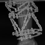
Part 10 (5 January 2016) – Complex Lower Leg Injury with Significant Injury to the Knee Joint and Extensor Mechanism

Part 10 – 5 January 2016
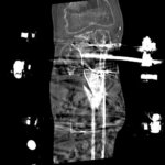
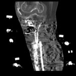
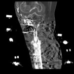
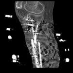
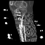
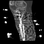
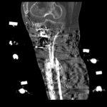
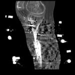
CT scan was done and showed very promising signs of healing. Not healed yet, but did not expect it anyway. I am very pleased with the results.
Otherwise pin sites still OK, no signs of infection. Walking slightly better but not as I would like it to be. Created an additional prescription for TSF to translate the distal tibia medially, but keep slight external rotation of the foot.
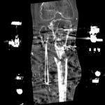
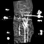
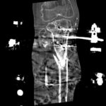
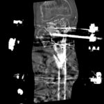
Below you can see sagittal and coronal reconstruction CT images, where good calus formation can be seen. Also you can see how we really managed to “shove” the proximal fragment into the mid fragment’s medullary canal. It really worked. You can also see some cement left in the lateral condyle which we were aware of from the beginning.
Plan:
- Complete the TSF prescription
- Continue mobilising
- Wait for consolidation of the regenerate and for the fracture to heal

Author: Matija Krkovic
Website: https://www.limbreconstructions.com/
