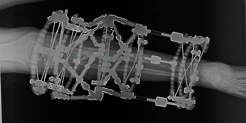
Part 8 (24 November 2015) – Complex Lower Leg Injury with Significant Injury to the Knee Joint and Extensor Mechanism

Part 8 – 24 November 2015
No major problems. Majority of pin sites dry with no signs of infection. Patient still not walking comfortable with one crutch due to the limited ankle dorsiflexion. Can only manage shorter distances with one crutch. Around 10-15 degrees flexion contracture in the right knee. Swelling of the leg significantly improved.
X-rays confirmed that the proximal fracture is healing with good callus formation and the distal regenerate is gaining in quality. So far as planned. There is clinically some valgus in his knee and X-ray confirms it (around 8-10 degrees). Question is whether to accept it or not?
I am personally more inclined to correct the alignment at the level of corticotomy using struts. This would represent third level of struts on one leg. It does sound complicated but certainly doable. To increase the stability will add a blind ring to the construct (to keep struts short).
Plan:
- CT scan through the proximal fracture to assess the union
- Exchange clickers with struts to correct the valgus while we still can
- Further follow up in 6 weeks

Author: Matija Krkovic
Website: https://www.limbreconstructions.com/