Limb Reconstructions > Blog > Clinical Blog > Complex Lower Leg Injury > Part 17 (4 October 2016) – Complex Lower Leg Injury with Significant Injury to the Knee Joint and Extensor Mechanism
Part 17 (4 October 2016) – Complex Lower Leg Injury with Significant Injury to the Knee Joint and Extensor Mechanism

Part 17 – 4 October 2016
Pt is still walking with crutches, shorter distances unaided. No further changes in lower leg alignment. At the moment there is 10-14 deg of valgus. Borderline deformity but we will accept it for now.
Pin sites still clean, no sign of instability.
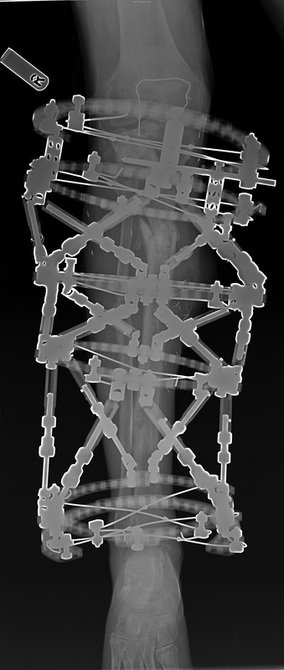
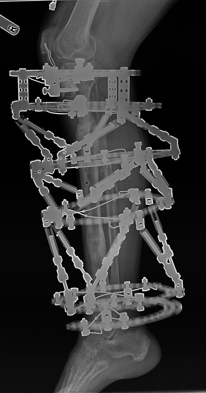
X-rays show further progress towards union in the proximal fracture when regenerate is well matured.
X-rays also show that fibula has been left in place and did not travel together with the distal tibia. I don’t know why I did not spot it earlier. Surprisingly there is no instability of the ankle joint neither any pain coming from the ankle joint.


Plan:
- CT scan requested
- Follow up in six weeks
bone healing CT scan external fixation fracture healing gait improvement limb reconstruction orthopedic surgery physiotherapy post-surgical recovery tibial alignment valgus deformity

Author: Matija Krkovic
Website: https://www.limbreconstructions.com/
I am a consultant orthopaedic trauma surgeon working at Addenbrooke's hospital, Cambridge University Hospitals NHS Foundation Trust.